Cochlear Implants
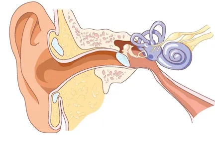
Anatomy of the ear
Sound waves travel through the external ear canal, vibrate the tympanic membrane (eardrum), and are transmitted through 3 middle ear bones. The stapes (a horseshoe-shaped bone) vibrates the oval window, transmitting waves to endolymphatic fluid inside the cochlea. Inside the cochlea, stereocilia (hair cells) on the basement membrane pivot, which transmits electrical signals to the auditory nerve.
Image from M. Lenoir, "Audition Promenade Round the Cochlea" website.
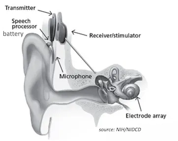
Cochlear implants
The cochlear implant is the most common neural implant and can allow hearing in those with congenital or acquired hearing loss. Greater than 1 million worldwide cochlear implants have been implanted. The cochlear implant requires use of an externally placed microphone, which makes it difficult to use in all situations, susceptible to external noise from wind or head coverings, and impacts the cosmetic aspect of the device.
Image from NIH/NIDCD
Implantable microphone
With colleagues from Mass Eye and Ear, MIT, and Columbia, the Fowler lab is developing an implantable microphone with the goal of integrating it into a totally implantable cochlear implant, eliminating the need for an external component.
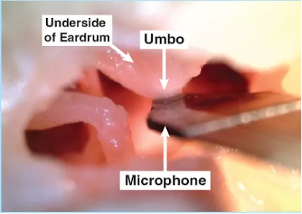
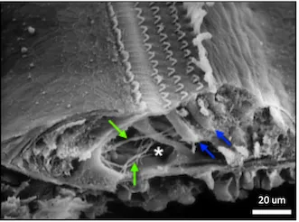
Sensor design
This microphone contacts the umbo, the portion of the manubrium (arm) of the malleus which vibrates along with the tympanic membrane. Benefits to this design include natural amplification from the ear canal. The microphone is a sensor with dual layers of PVDF, a piezoelectric material, coupled to titanium electrodes.
Future studies
Future steps include conducting a live study using sheep as a large animal model. In addition, the lab is developing a fixation device to stabilize the microphone in the middle ear.